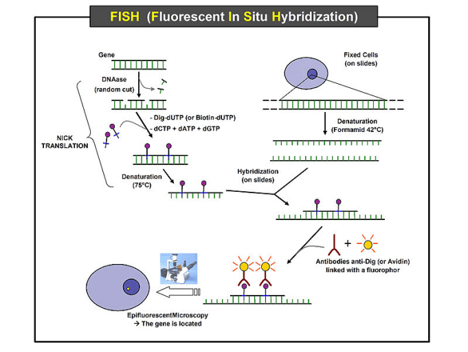
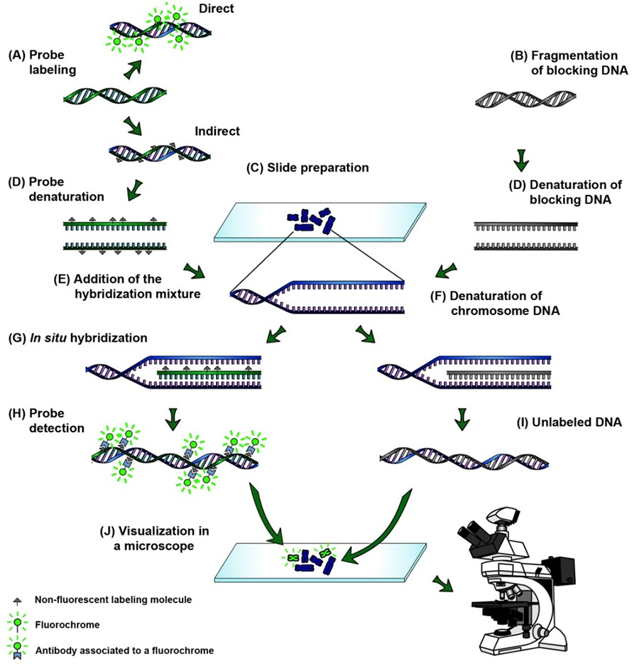
Labeled ssDNA or ssRNA probes are used to hybridize with in vivo mRNA or DNA that is denatured to become ssDNA prior to hybridization. This labeled RNA or DNA probe can then be detected by using an antibody to detect the label on the probe.

Fluorescence In Situ Hybridization Fish Protocol Creative Biomart
Ideal probes are the entire length of your gene.

. In situ hybridization is a cytological method of mappinga probe is complemetay to a chromosomal sequence is used to locate the gene microxcopically within a mixture of many different chromosomes. In this chapter we provide a systematic overview of the published guidelines and validation procedures for fluorescence in situ hybridization FISH probes for clinical diagnostic use. Oligonucleotide probes are usually 2050 bases in length.
I prefer targeting the 3 end of the gene and try to anchor at least 12 or 23 of that probes length to be in the 3UTR. Output probes must be filtered through. FISH is a molecular cytogenetic technique that uses fluorescent probes to detect specific DNA sequences on the chromosome.
The longer the probe the higher the hybridization temperature can be used If oligonucleotide probes are used the hybridization temperature is low the formamide concentration low the salt concentration high If long probes DNA or RNA are used the higher the temperature the. In situ hybridization ISH is a type of hybridization that uses a labeled complementary DNA RNA or modified nucleic acids strand ie probe to localize a specific DNA or RNA sequence in a portion or section of tissue in situ or if the tissue is small. In situ hybridization protocolGeneral procedure and tips for in situ hybridization using antibody detection.
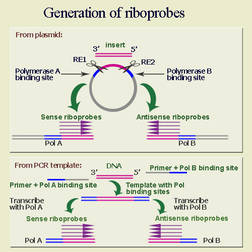
Riboprobes transcribedinoppositeorienta- tions serve this purpose for detection of mRNA by the. Fluorescence in situ hybridization FISH can be used to visualize spatial locations of DNA regions and RNA molecules in a sequence-specific manner. When more than one probe is used the order of genes along a particiular chromosome can be.
The probe could be a. Templates must be linearized before making a probe though PCR templates can also be used. In situ hybridization The methods used to localize mRNA or single-stranded ss DNA at the tissue or cellular level.
In situ hybridization probes are labeled DNA or RNA oligonucleotides that are complementary to genes or genomic regions of interest. There are essentially four types of probe that can be used in performing in situ hybridization. Today most in situ hybridization procedures use fluorescent probes to detect DNA sequences and the process is commonly referred to as FISH fluorescence in situ hybridization.
A labeled RNA or DNA probe can be used to hybridize to a known target mRNA or DNA sequence within a sample. Dabbs MD in Diagnostic Immunohistochemistry 2019 HER2 Fluorescence In Situ Hybridization and Other In Situ Hybridization Assays. In situ hybridization of wild type Drosophila embryos at different developmental stages for the RNA from a gene called hunchback.
The probe has a chemical or radioactive label attached to it so that its binding can be observed. Probes should be at least 1 kb. Modern methods using short oligonucleotide probes have eliminated the use of long RNA probes but the requirement of many short probes has limited the application to long transcripts and incur high cost when proprietary reagents are used.
Targets include chromosomal loci for DNA probes or transcribed RNA usually mRNA for. They do not hybridize as strongly to target mRNA molecules compared to RNA probes so formaldehyde should not be used in the post hybridization washes. When used in in situ hybridization RNA probes are _____ to the samples RNA.
Use Design primers and select forward primer. The probes can therefore be used to detect. A labeled RNA or DNA probe can be used to hybridize to a known target mRNA or DNA sequence within a sample.
Methodological and theoretical aspects of ISH are first considered with regard to tissue fixation probe selection and preparation various types of labeling reactions and aspects of hybridization conditions as they relate to hybridization protocols. IN SITU HYBRIDIZATION Control Probes Proper in situ hybridization technique demands the in- clusion of control probes to demonstrate that binding is specific and not due to vector sequence or to non-specific hybridization. Rescence in situ hybridization FISH probes extensively to make clinical diagnoses and under United States law these laboratories must validate their FISH probes before use.
Definition Procedures Analysis Worksheet 1. In case ofHER2 FISH a HER2 probe is used to identifyHER2 gene amplification. As far as designing in situ probes I typically TA clone 1-15kb probes.
In situ hybridization is a laboratory technique in which a single-stranded DNA or RNA sequence called a probe is allowed to form complementary base pairs with DNA or RNA present in a tissue or chromosome sample. In situ hybridization indicates the localization of gene expression in their cellular environment. The technique of in situ hybridization ISH is presented with special reference to its use in the practice of pathology.
In Situ Hybridization Probes. In situ hybridization indicates the localization of gene expression in their cellular environment. FISH probes-which are classified as molecular probes or analyte-specific reagents ASRs-have been extensively used in vitro for both clinical diagnosis and research.
They are produced synthetically by an automated chemical synthesis employing a specific DNA nucleotide sequence of your choice. In this type of histochemical application cell and tissue samples are incubated with probes which localize to sites within the cell. This labeled RNA or DNA probe can.
Using Geneious select 200-250 bp at the beginning of your gene sequence. If the gene is 2 kb the probe can be a 1-2 kb fragment of the gene. Print In Situ Hybridization ISH.
DNA probes DNA probes provide high sensitivity for in situ hybridization. In situ hybridization is widely used for detection of cellular RNA. Kinetics of hybridization Probe length.
It can be used to cytologically map the location of a gene sequence.
Fluorescent In Situ Hybridization Flow Chart The Samples Are Fixed Download Scientific Diagram

In Situ Hybridization Ish And Fluorescence In Situ Hybridization Fish Creative Diagnostics

0 Comments